Introduction


      We sample the world through our photoreceptors (rods and cones ) which are located at the back of the retina. The resolution at which we can resolve the world is related to the spatial distribution of the cones (the rods sample the retinal image much more finely than the cones but the signals from many rods converge to a single neuron so this fine resolution is lost). Three types of cones - S (blue), M (green) and L (red) - are found in the primate retina. They have their peak sensitivity respectively in the Short, Middle and Long wavelengths of the visible spectrum.


      In (DeMonasterio, 1981), de Monasterio looked in fluorescence microscopy at the monkey retina using a fluorescent dye which stains the S-cones. He found that the "cones form an approximately regular array" whose separation increases toward the periphery of the retina. Assuming such a regular distribution, several aliasing experiments have been done to estimate the spacing between the S-cones within the fovea in human (Williams, 1983). Outside the fovea, the irregularities of the sampling mosaic have been argued to be a way to alleviate aliasing (Yellott, 1983).


      However, in (Curcio, 1991), S-cones even within the fovea in human were found to be sparse and irregularly spaced. In (Roorda, 1999), Roorda used adaptive optics and retinal densitometry to look at the arrangement of S, M and L cones in vivo. He could not distinguish the distribution of the S cones from random and found that the M and L cones were either randomly distributed or aggregated. Later work (Roorda, 2001) indicates a random distribution of S, M and L cones in human, but recent studies "suggest that the assignment of L and M pigment, although highly irregular, is not a completely random process" (Hofer, 2005).


      If indeed, the distribution is (almost) random, what does this randomness mean? Can we generalize to the fovea what people originally thought of the periphery, namely that irregularities are actually useful?


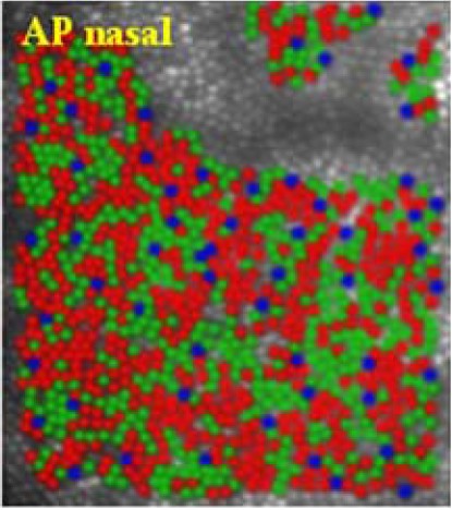
Figure 1:  False color images showing the arrangement of L (red), M (green), and S (blue) cones in the retinas of different human subjects (From Hofer, 2005).


Home Introduction The Visual System Compressive Sensing Single-Pixel Imaging Conclusions References